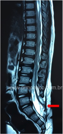
Tethered spinal cord is a common condition in people who have so-called hidden dysraphism in the spine or in those who presented myelomeningocele and underwent surgical treatment as a baby.
The normal spinal cord is loose within the vertebral canal, surrounded by the cerebrospinal fluid (CSF), moving freely according to body movement or growth. When the spinal cord shows adhesion, especially in the lumbar or sacral region, it is literally trapped, susceptible to micro-impact injuries (since it does not move freely within the cerebrospinal fluid). When a child has spinal cord attached, as growth occurs, it undergoes a stretch and may give the following symptoms:
1) Urinary dysfunction (neurogenic bladder)
2) Intestinal dysfunction (neurogenic intestine)
3) Spasticity in lower limbs
4) Pain
5) Worsening of scoliosis or other deformities in the spine
6) Foot deformities
7) Worsening gait or weakness in lower limbs
The spinal cord prey can occur in those who did not operate myelomeningocele as a baby, when associated with so-called hidden dysraphisms. They are: lipomielomeningocele, spina bifida (diastematomyelia), adhesion of terminal filum, spinal cord dermoid cyst, congenital dermal sinus, presence of lumbar or sacral dimple (like a lumbar or sacral lumbar). Cutaneous stigmas may be present in occult dysraphisms, such as small tufts of hairs in the lumbar or sacral region, blemishes in the region or roughness in the skin.
When the tethered spinal cord has already been identified by imaging examination, but the person is asymptomatic, no surgical treatment is required. When there are progressive symptoms, the treatment consists of a microsurgery for the release of the spinal cord prey. Surgery is performed through microscopy with access generally to a scarred region and fibroses that may make it difficult to identify what is neural tissue. The use of intraoperative neurophysiological monitoring is recommended to minimize or avoid neural damage. Duramater is often absent, and CSF leakage is a risk.
The expected result with the surgical procedure is mainly improvement in spasticity and pain. The symptoms of deformities may improve, but not necessarily reverse. The surgery works more to prevent a progression of deformities than to promote a regression of the same. Complementary orthopedic surgeries are usually needed.

Magnetic resonance imaging (MRI) showing a medulla attached at the sacral level (red arrow), in a region where there is failure of the spinal closure (patient with myelomeningocele). Dr Bernardo de Monaco.
For more information, search a specialist neurosurgeon.

